






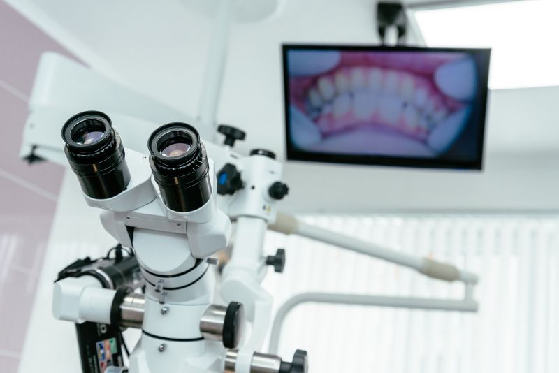
When is a microscope used in root canal treatment?
Everyone understands that sometimes the quality of the work depends not only on the talent of the specialist doing it but also on the materials and technique used. This is especially relevant when it comes to oral health. Without a professional microscope it's impossible to carry out quality root canal treatment, because causes invisible to the naked eye not infrequently lead to very serious tooth-health problems that require additional expense and time.
In dentistry, a microscope is most often used precisely in root canal treatment. It's needed here because dental canals are very fine: their length varies from 10 to 20 mm, and at the narrowest points the working endodontic area can be just 0.3–0.6 mm. Any imprecise mechanical action in the root, or an instrument carelessly inserted into it, can cause the tooth root to split. A microscope also helps find dental canals and micro-damage to the tooth roots.
Another benefit of a microscope is that it enlarges the field of view when working in the canals with ultrasound, when filling materials or other foreign bodies that can cause inflammation need to be removed from the dental canals.
A microscope is also used when the inner tissues of a tooth have to be treated, to endodontically open the cavities and pulp of an infected tooth, clean out inflammations and remove the tooth nerve.
So when choosing a dental clinic, you should assess whether it has a microscope the dentist can use during any procedure. After all, no one doubts that diagnosing a problem with the help of a microscope and eliminating it straight away is far better than corrections later.
We're glad that at AND klinika we have an advanced microscope our dentists can always call on, so that by entrusting your dental treatment to us, you can be sure everything will be done flawlessly and precisely.